3D Printing in Anatomy Teaching
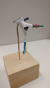
 Several members of staff have been working on a model of the pterygopalatine fossa – this an anatomical space within the skull that is conceptually very difficult to understand. Using imaging software and a CT scan of the skull, we created a 3D render of the space. This enabled us to produce eight identical models with a 3D printer. The models were then painted and mounted on stands.
The models were used in the dissection room to enhance 3rd year head and neck anatomy teaching. Dr Shivani Parihar and Dr Ross Bannon gave a poster presentation about our work at the ENT Scotland winter meeting on 18/11/16 and received an honourable mention at the prize-giving ceremony!
Many people have contributed to this project:
Ross Bannon
Shivani Parihar
Yiannis Skarparis
Ian Gordon
Murray Coutts
Ian Parkin
Fraser Chisholm

Dr Shivani Parihar and Dr Ross Bannon with their poster at the ENT Scotland Winter meeting